Gallium »
PDB 1cfw-9ohs »
1efd »
Gallium in PDB 1efd: Periplasmic Ferric Siderophore Binding Protein Fhud Complexed with Gallichrome
Protein crystallography data
The structure of Periplasmic Ferric Siderophore Binding Protein Fhud Complexed with Gallichrome, PDB code: 1efd
was solved by
T.E.Clarke,
S.-Y.Ku,
D.R.Dougan,
H.J.Vogel,
L.W.Tari,
with X-Ray Crystallography technique. A brief refinement statistics is given in the table below:
| Resolution Low / High (Å) | 20.00 / 1.90 |
| Space group | P 63 |
| Cell size a, b, c (Å), α, β, γ (°) | 86.540, 86.540, 91.770, 90.00, 90.00, 120.00 |
| R / Rfree (%) | 21.8 / 25 |
Gallium Binding Sites:
The binding sites of Gallium atom in the Periplasmic Ferric Siderophore Binding Protein Fhud Complexed with Gallichrome
(pdb code 1efd). This binding sites where shown within
5.0 Angstroms radius around Gallium atom.
In total only one binding site of Gallium was determined in the Periplasmic Ferric Siderophore Binding Protein Fhud Complexed with Gallichrome, PDB code: 1efd:
In total only one binding site of Gallium was determined in the Periplasmic Ferric Siderophore Binding Protein Fhud Complexed with Gallichrome, PDB code: 1efd:
Gallium binding site 1 out of 1 in 1efd
Go back to
Gallium binding site 1 out
of 1 in the Periplasmic Ferric Siderophore Binding Protein Fhud Complexed with Gallichrome
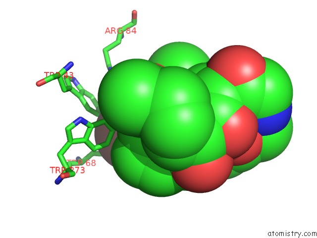
Mono view
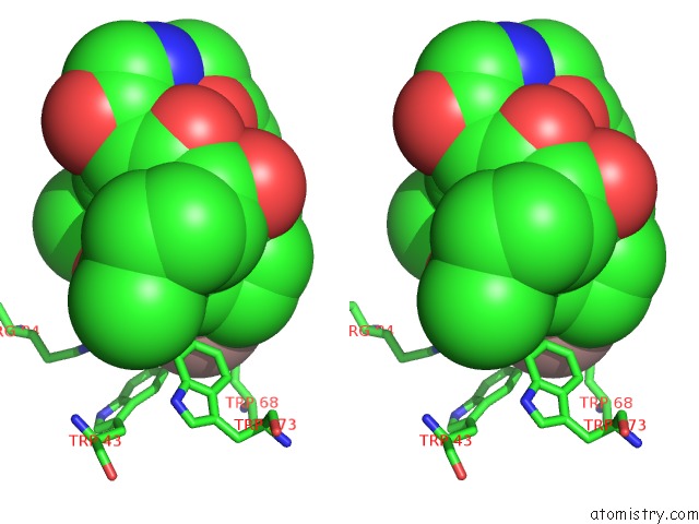
Stereo pair view
Mono view
Stereo pair view
A full contact list of Gallium with other atoms in the Ga binding
site number 1 of Periplasmic Ferric Siderophore Binding Protein Fhud Complexed with Gallichrome within 5.0Å range:
|
Reference:
T.E.Clarke,
S.Y.Ku,
D.R.Dougan,
H.J.Vogel,
L.W.Tari.
The Structure of the Ferric Siderophore Binding Protein Fhud Complexed with Gallichrome. Nat.Struct.Biol. V. 7 287 2000.
ISSN: ISSN 1072-8368
PubMed: 10742172
DOI: 10.1038/74048
Page generated: Fri Aug 8 08:23:17 2025
ISSN: ISSN 1072-8368
PubMed: 10742172
DOI: 10.1038/74048
Last articles
I in 1GZAI in 1GWD
I in 1GUL
I in 1GTE
I in 1GTH
I in 1GJD
I in 1F3M
I in 1FZ9
I in 1GA5
I in 1F86