Gallium »
PDB 1cfw-9ohs »
5auk »
Gallium in PDB 5auk: Crystal Structure of the Ga-Substituted Ferredoxin
Protein crystallography data
The structure of Crystal Structure of the Ga-Substituted Ferredoxin, PDB code: 5auk
was solved by
G.Kurisu,
N.Muraki,
M.Taya,
T.Hase,
with X-Ray Crystallography technique. A brief refinement statistics is given in the table below:
| Resolution Low / High (Å) | 27.56 / 1.62 |
| Space group | P 61 2 2 |
| Cell size a, b, c (Å), α, β, γ (°) | 31.942, 31.942, 320.078, 90.00, 90.00, 120.00 |
| R / Rfree (%) | 19.8 / 21.9 |
Gallium Binding Sites:
The binding sites of Gallium atom in the Crystal Structure of the Ga-Substituted Ferredoxin
(pdb code 5auk). This binding sites where shown within
5.0 Angstroms radius around Gallium atom.
In total 2 binding sites of Gallium where determined in the Crystal Structure of the Ga-Substituted Ferredoxin, PDB code: 5auk:
Jump to Gallium binding site number: 1; 2;
In total 2 binding sites of Gallium where determined in the Crystal Structure of the Ga-Substituted Ferredoxin, PDB code: 5auk:
Jump to Gallium binding site number: 1; 2;
Gallium binding site 1 out of 2 in 5auk
Go back to
Gallium binding site 1 out
of 2 in the Crystal Structure of the Ga-Substituted Ferredoxin
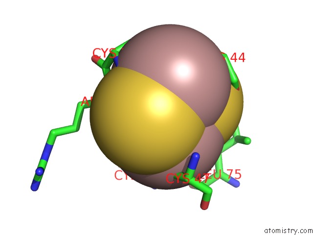
Mono view

Stereo pair view
Mono view
Stereo pair view
A full contact list of Gallium with other atoms in the Ga binding
site number 1 of Crystal Structure of the Ga-Substituted Ferredoxin within 5.0Å range:
|
Gallium binding site 2 out of 2 in 5auk
Go back to
Gallium binding site 2 out
of 2 in the Crystal Structure of the Ga-Substituted Ferredoxin
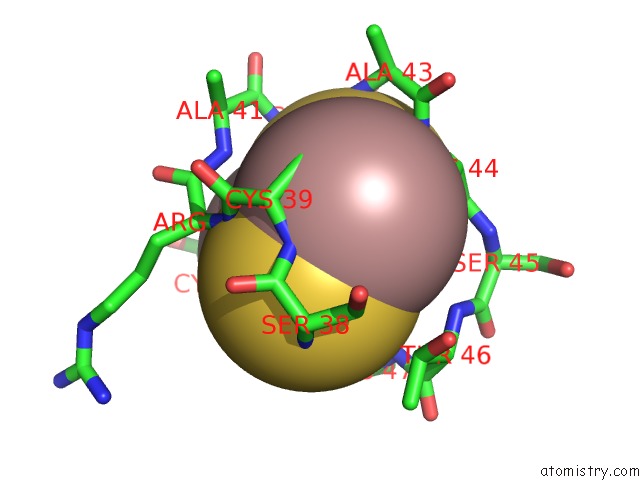
Mono view
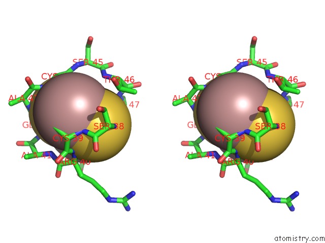
Stereo pair view
Mono view
Stereo pair view
A full contact list of Gallium with other atoms in the Ga binding
site number 2 of Crystal Structure of the Ga-Substituted Ferredoxin within 5.0Å range:
|
Reference:
R.Mutoh,
N.Muraki,
K.Shinmura,
H.Kubota-Kawai,
Y.H.Lee,
M.M.Nowaczyk,
M.Rogner,
T.Hase,
T.Ikegami,
G.Kurisu.
X-Ray Structure and Nuclear Magnetic Resonance Analysis of the Interaction Sites of the Ga-Substituted Cyanobacterial Ferredoxin Biochemistry V. 54 6052 2015.
ISSN: ISSN 0006-2960
PubMed: 26348494
DOI: 10.1021/ACS.BIOCHEM.5B00601
Page generated: Fri Aug 8 08:23:51 2025
ISSN: ISSN 0006-2960
PubMed: 26348494
DOI: 10.1021/ACS.BIOCHEM.5B00601
Last articles
I in 1GZAI in 1GWD
I in 1GUL
I in 1GTE
I in 1GTH
I in 1GJD
I in 1F3M
I in 1FZ9
I in 1GA5
I in 1F86